How do you check anterior tibialis?

Noah Mitchell
Published Jan 17, 2026
Place your resistance hand on the medial side of the distal foot. Resist the client from dorsiflexing and inverting the foot. Look the distal tendon of the tibialis anterior on the medial side of the ankle joint and foot; it is usually visible. Palpate the distal tendon by strumming perpendicular across it.
How do you test for tibialis anterior tendonitis?
Assessment tests
Pain at the front of the ankle on either of these tests may indicate tibialis anterior tendon pain. An ultrasound scan or MRI scan may be used to confirm the diagnosis and rule out a strain or tear of the tendon.
How do you test your tibialis posterior?
Manual muscle testing of the tibialis posterior is performed by placing the foot in an everted, plantar flexed position and the patient is asked to invert the foot. Weakness or pain during contraction of an injured tibialis posterior muscle is characteristic.
How does tibialis anterior feel?
Symptoms of Anterior Tibialis Tendinitis
Pain when lifting foot or toes, swelling, feeling of ankle weakness, or tenderness when palpating the tendon. The pain can increase with activity, most commonly with walking or running uphill or downhill. Overuse is often the most common cause of anterior tibialis tendinitis.
What Innervates tibialis anterior?
The tibialis anterior is one of four muscles in the anterior compartment of the leg. The others include extensor digitorum longus (EDL), extensor hallucis longus (EHL), and fibularis tertius. The deep peroneal nerve innervates all muscles and is perfused by the anterior tibial artery.
24 related questions foundHow do you train anterior tibialis?
Tibialis Anterior Strengthening Exercises
- Seated Toe Raises. Sit on a chair with your feet in front of you. Slowly raise your toes off of the floor. ...
- Wall Toe Raises. Stand 12 inches away with your back towards the wall with feet hip width apart. ...
- Heel Walk. Stand on both feet hip width apart with no shoes on.
What causes a sore tibialis anterior?
The anterior tibial tendon lies on the inner-front of the ankle. The muscle and tendon work together to flex the foot upwards. This condition occurs when the tendon is inflamed from overuse or traumatic ankle injury. If left untreated, the tendon can rupture and is very difficult to treat.
Why is my tibialis anterior sore?
Tibialis anterior pain is usually felt during or after activities that are stressful for the muscle such as excessive walking, running, or hiking (especially up and down hills or on hard surfaces), kicking with pointed toes (soccer), or wearing tight shoes. Pain may be felt in the ankle, foot, or shin.
Can you walk with anterior tibial tendonitis?
The tibialis anterior tendon attaches a muscle in the front of your shin to the front of your foot. A tear of this tendon may cause pain and difficulty performing normal activities like walking and running.
Where do you palpate tibialis posterior?
The posterior tibial pulse can be felt behind and below the medial malleolus. Gently flex the knee and feel for the popliteal pulse by deep palpation in midline in popliteal fossa.
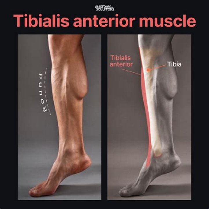
Where is the tibialis anterior located?
The Tibialis anterior (Tibialis anticus) is situated on the lateral side of the tibia; it is thick and fleshy above, tendinous below. The fibers run vertically downward, and end in a tendon, which is apparent on the anterior surface of the muscle at the lower third of the leg.
How long does it take for tibialis anterior to heal?
The healing time for anterior tibialis tendon repair will take up to 8-12 weeks but restoration of function and ability to accept full activity, load and stress can take up to one year.
What is tibialis anterior strain?
Anterior tibialis tendonitis is an injury of the anterior tibialis tendon in the front of the ankle where it meets the foot. The tendon is important in pulling the foot up (dorsiflexion), lifting the foot off the ground while running, and in turning the foot inward (inversion).
Do calf raises work tibialis anterior?
With a slight adjustment of your body position, you can use a standing calf raise machine to target the tibialis anterior muscle.
What are the symptoms of a damaged tibial nerve?
Tibial nerve dysfunction occurs when there is damage to the tibial nerve. Symptoms can include numbness, pain, tingling, and weakness of the knee or foot. The tibial nerve is commonly injured by fractures or other injury to the back of the knee or the lower leg.
How do you fix anterior tibialis pain?
Focus on reducing stress on the tibialis anterior by wearing shoes with a lower heel and sticking to softer surfaces when you run, and increase the tendon and muscle's strength by doing heel walks and wall toe raises. Optionally, you can try kinesiology taping and compression wear to help speed your recovery.
Is the anterior tibialis a flexor or extensor?
The tibialis anterior muscle is flexor, inverter (in addition to posterior tibial muscle) and adductor (in addition to the long extensor of hallux) of the foot. It also plays a role in suspension of the arch and controls supination of the rearfoot [10].
Where do you palpate anterior tibial artery?
The anterior tibial artery pulse can be palpated near the origin of the dorsalis pedis artery on the dorsum of the foot lateral to the extensor hallucis longus tendon.
What Innervates the posterior tibialis?
Innervation. Tibialis posterior is innervated by the tibial nerve which arises from the L4 and L5 spinal nerves. The tibial nerve is the larger of the two branches of the sciatic nerve.
Which muscles Dorsiflex the foot at the ankle?
The tibialis anterior muscle, found in the anterior compartment of the leg, is the primary muscle that facilitates dorsiflexion of the ankle joint. The peroneus longus and Peroneus Brevis muscles, found in the lateral compartment of the leg, function to facilitate eversion of the ankle joint.
How do you test for ankle inversion?
Therapist is sitting on a stool in front of patient. One hand stabilizes the ankle just above the malleoli while other hand provides resistance around the dorsum and lateral border of the forefoot. Resistance is directed toward inversion and slight dorsiflexion. Patient actively turns the foot down and out.