What are perforating Sharpey's fibers and what is their function?

Sarah Smith
Published Jan 07, 2026
In the skull the main function of Sharpey's fibres is to bind the cranial bones in a firm but moveable manner; they are most numerous in areas where the bones are subjected to the greatest forces of separation. In the spine, similar fibres join the intervertebral disc to the adjacent vertebrae.
What is the function of the perforating fibers of the skeletal system?
Perforating fibers--- bundles of collagen fibers that extend into the bone matrix --- secure the periosteum to the underlying bone. the periosteum also provides anchoring points for tendons and ligaments.
What are perforating fibers in bone?
n. Any of the bundles of collagen fibers that pass into the outer circumferential lamellae of bone or into the cementum of teeth.
What are Sharpey's fibers in cementum?
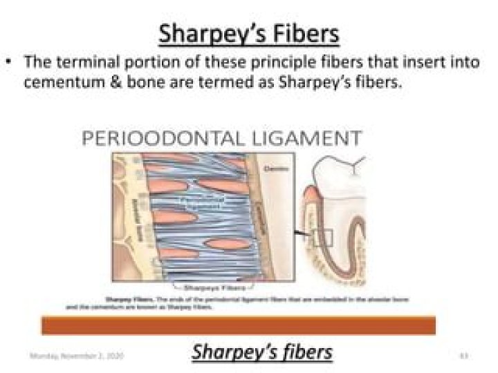
Sharpey's fibers (perforating fibers) are portions of the principal collagenous fibers of the periodontal ligament embedded in the cementum and alveolar bone that attach the tooth to the alveolus.
What are Sharpey's fibers histology?
Sharpey's Fibers of Cementum
Collagen fibers formed by fibroblasts of the PDL (E) forming the principal fiber bundles, are partly incorporated into the cementum (A). They are referred to as extrinsic or Sharpey's fibers (F). Note that the terminology corresponds to that used in bone tissue.
35 related questions foundWhere is the perforating canal located?
At the base of individual osteons are perforating canals (also called Volkmann's canals), which are empty spaces that allow blood vessels, lymph vessels, and nerves to travel across bone, linking up with the vessels and nerves in the central canals.
What two structures do the perforating fibers connect?
The periosteum attaches to the bone via distinct collagen bundles, which are perforating fibres, that connect calcified bone matrices and other organs.
What is cellular cementum?
Cellular cementum contains cells and is the medium of attachment of collagen fibres to the alveolar bone. It is also responsible for minor repair of any resorption by continued deposition to keep the attachment apparatus intact. Acellular cementum does not contain cells and has a main purpose of adaptive function.
How is cellular cementum formed?
The development of acellular cementum seems to be associated with secretion of enamel-related proteins by cells of the epithelial root sheath. Formation of the matrix for cellular cementum appears to be induced by exposure of the inner layer of the epithelial root sheath to the mesenchymal cells in the dental follicle.
What is fenestration and dehiscence?
Fenestration is the condition, in which the bony coverage of the root surface is lost, and the root surface is only covered by the periosteum and gingiva. In such lesions, marginal bone is intact. When this bone defect spreads toward the marginal bone, it is called dehiscence.[1]
What is a perforating canal?
Perforating canals provide channels that allow the blood vessels that run through the central canals to connect to the blood vessels in the periosteum that surrounds the bone.
Where are Sharpey's fibers attached?
Bone consists of highly calcified, intercellular bone matrix, and three types of cells—osteocytes, osteoblasts, and osteoclasts. The outer surface of bone is covered by periosteum, which is bound to bone by bundles of collagen fibers known as Sharpey's fibers, and the inner bone surface is lined with endosteum (Fig.
What is the function of Sharpey fibers quizlet?
Collagenous fibers (Sharpey fibers) that penetrate the bone anchor the inner layer of the periosteum to the bone. Sharpey fibers help hold or attach tendons and ligaments, not muscle, but to the periosteum of bones.
Which of the following is a function of synovial fluid quizlet?
What is the function of the synovial fluid? Synovial fluid moistens and lubricates the joints, as well as supplying nutrients to the articular cartilage.
What is the function of the medullary cavity?
The medullary cavity is the hollow part of bone that contains bone marrow. The bone marrow makes blood cells and stores fat.
Which is secreted by osteoblasts during bone deposition?
Osteoblasts secrete the extracellular matrix and deposit calcium, which hardens the matrix. The non-mineralized portion of the bone or osteoid continues to form around blood vessels, forming spongy bone. Connective tissue in the matrix differentiates into red bone marrow in the fetus.
Are dentin forming cells?
Odontoblasts are specialized cells that produce dentin and exhibit unique morphological characteristics; i.e., they extend cytoplasmic processes into dentinal tubules.
Is dentin vascular or avascular?
Dentin is a yellowish, somewhat elastic but mineralized avascular tissue that supports the enamel and encloses the pulp chamber.
Which of the following cell is responsible for Cementogenesis?
28.2.
Key cells in root formation are Hertwig's epithelial root sheath (HERS) cells. They appear to be involved in the initiation of the process, in root elongation, and in stimulating both cementogenesis and root dentinogenesis [28].
What is the alveolar process?
The alveolar process is the horizontal portion of the maxilla that holds the tooth roots. b. Alveoli for the tooth roots are present all along the alveolar process, except where these have been resorbed following the loss of teeth.
What is the function of cementum in tooth?
The main function of cementum is tooth support or tooth anchorage together with the principal fibers and alveolar bone.
What is dentin teeth?
Dentin. That part of the tooth that is beneath enamel and cementum. It contains microscopic tubules (small hollow tubes or canals). When dentin loses its protective covering (enamel), the tubules allow heat and cold or acidic or sticky foods to stimulate the nerves and cells inside the tooth, causing sensitivity.
Are tendons a muscle?
Overview. A tendon is a fibrous connective tissue that attaches muscle to bone. Tendons may also attach muscles to structures such as the eyeball.
What are the different types of bone cells and their functions?
There are three types of cells that contribute to bone homeostasis. Osteoblasts are bone-forming cell, osteoclasts resorb or break down bone, and osteocytes are mature bone cells. An equilibrium between osteoblasts and osteoclasts maintains bone tissue.
Are large phagocytic cells found in bone?
Osteoclasts are large multinucleated phagocytic cells derived from the macrophage-monocyte cell lineage (23). They migrate from bone marrow to a specific skeletal site.